Most of the things I am interested in most other people haven’t heard of and would have little interest in them if they had. So it is with diatoms. About half the people I mention them to have no idea what they are. They are very surprised when I tell them that they are estimated to produce about 20% of the oxygen we breathe – 50% when grouped with other phytoplankton. Through photosynthesis, diatoms drifting in the world’s oceans produce more oxygen and fix more carbon dioxide than all of the Earth’s forests combined. Since they form the foundational base of aquatic food webs our oceans would be devoid of fish without them. Those facts surprise almost anyone who can be bothered to listen to me prattle on about them. Their surprise intensifies when I tell them that they are algae that are partly made of glass and yet despite living in a glass shell, they can move. If I am honest and despite doing various courses on botany as an undergraduate, much of the above originally came to me as a surprise.
The glass shells or frustules of diatoms are often beautifully patterned with striae and dots so fine that they are used to test the performance of light microscopes. Using diatoms to check the resolving power of the optical train of a microscope was first suggested by J. D. Sollit and his collaborator Robert Harrison in 1841. Sollitt discovered that the incredibly fine, ultra-precise periodic structures on the diatom Pleurosigma hippocampus could only be resolved into clear lines or dots by high-quality microscope lenses. Recognizing their value for testing optical performance, he sent specimens to the world’s leading microscope makers of the time (including Smith, Ross, Powell & Lealand in London, and Nachet in Paris). His idea sparked what can only be described as an ‘optical arms-race’ as different manufacturers vied to produce objectives of ever higher numerical aperture and thus resolving power. Sollitt claimed to have been able to resolve the 130,000 lines per inch on the frustules of Amphipleura pellucida. This is equivalent to more than 5000 lines per millimetre and since the distance between the lines is less than the wavelength of blue light, there was some debate as to whether or not this was even possible with the dry low numerical aperture lenses he would have had available to him. What is in no doubt is that his idea of using diatoms as test objects was one of the driving forces behind the rapid advance in microscope optics seen in the last century.
Sollitt was a classic Victorian gentleman amateur who was headmaster of the Hull Grammar School in Yorkshire. He taught Latin and mathematics but his true passion was for natural philosophy and he became one of the most meticulous and fiercely competitive microscopists of the mid-19th century. While Sollitt was the master of optics, mathematical computation, and recording data, his colleague Robert Harrison was an expert at locating and collecting rare species of diatom, Their partnership turned 19th century Hull into an unlikely centre for breakthroughs in optics and microscopy. Sollit utilised blue glass and oblique illumination to enable him to push microscopes to the limits of their resolving power. Before Sollit popularized test diatoms, lens makers optimized their microscope lenses to eliminate chromatic and spherical aberration. Sollitt showed that a lens could be perfect in those respects but fail to resolve detail if it lacked a wide numerical aperture. His public challenges forced optical manufacturers to radically widen the “angular aperture” or what we now call the numerical aperture (NA), of their objectives. Sollitt would test the lenses made by leading manufacturers against the finest lines to be found on the diatoms collected by Harrison. On this basis, the maker’s claims would either be debunked or validated. The debate concerning Sollit’s claim to have resolved more than 5,000 lines per millimetre led to suggestions that he was lying. The arguments continued for decades. However, the modern consensus is that he wasn’t lying but rather that what he saw was an optical illusion called diffraction ghosting; interference patterns produced when light passes through narrow slits producing the illusion of twice as many lines as there actually are.
The ornate glass houses that contain the cytoplasm and organelles of diatoms or frustules, are made of two parts that fit together rather like the top and bottom halves of a close fitting tin and its lid. Thus, sex is a bit of a problem. Diatoms usually reproduce asexually; the smaller bottom half becomes the top half of a new and thus smaller diatom, while the top half generates a new bottom half resulting in a new diatom of the same size as the original one. The result of continued asexual reproduction is that the population of diatoms gets progressively smaller! When a critical size limit is reached diatoms reproduce sexually producing gametes that fuse to generate an auxospore. The auxospore like the parent diatoms is usually photosynthetic and depends on light to grow. However, in some cases it can be formed without any further supply of energy. A key feature of the auxospore is that it is distensible and after reaching its maximum size it turns into a new diatom with a rigid frustule, that is significantly larger than its parent cells. Thus, sexual reproduction within the diatom population increases its average size. I can’t think of any other example of sexual reproduction being triggered by a reduction in the size of the parents but, diatoms are really weird.
Diatoms are to be found almost everywhere that there is water; even among the leaves of mosses. Most other algae and plants generally, can only move through growth or because they get carried by wind or water. However, many diatoms are capable of active movement. Motile diatoms have a specialized, elongated slit-like groove or channel in their glass shells called a raphe through which they can secret sticky mucus a bit like the slime exuded by snails but in the case of diatoms the mucus is an adhesive via which the cell can pull itself forward. I am always surprised at just how fast diatoms can move. The fastest observed speed is about 35 um/s which may not seem fast until you consider that diatoms don’t have any appendages. In a remarkable example of the evolutionary conservation of the mechanisms of movement, diatoms rely on actin and myosin for their motion – the very same molecules found in our own muscles.
While observing living diatoms reveals much about their nature, the presence of chlorophyll and carotenoids gives them a golden brown colour that masks the ornate structure of their frustules. Observation of the shells of living diatoms is made all the more difficult because glass has a refractive index close to that of water. In order to make the architecture of the frustule visible it is necessary to both remove the pigment they contain and use techniques that will either enhance the contrast of the frustule or create a larger difference in refractive index than can be provided by aqueous media.
I have collected diatoms adhering to stones in rivers in Japan and France. I retrieve them by scrubbing the stones with a toothbrush and repeatedly tipping water over the surface of the stone. Inevitably, such samples are contaminated with other algae, minerals and other assorted ‘gunge’. The pigment in the diatoms and these contaminates have to be removed. In the old days Victorian microscopists would do this by boiling their samples in a mixture of concentrated nitric and sulphuric acids. Once cleaned with these chemicals and washed free of them, the diatoms were mounted on a slide either in air or within a high refractive index medium of which there were several but that could only be created using other highly toxic chemicals. Unfortunately (?), strong acids are no longer sold to the general public nor are the chemicals used to create high refractive index mountants. Fortunately, there are somewhat less toxic ways of freeing the shells of diatoms from other gunk and it’s possible to buy suitable mountants from suppliers of reagents for microscopy. I will not go into the details of my techniques here but rather I will provide the recipes in another blog post. What I will say is that most of the things I tried – boiling in bleach, treatment with hydrogen peroxide etc. didn’t work! It was a long road filled with many disappointments but eventually as I hope the pictures will show, I enjoyed a little success.
The next steps in my diatom journey will be to try to ‘arrange diatoms’. Diatom arrangement is an art-form that dates back to the early days of microscopy. In essence it involves picking diatoms up and moving them around to create attractive mosaics or images. I have built the apparatus for doing this but so far I have had close to zero success. For anyone interested in what such ‘arrangements’ can look like I would urge you to watch this video featuring the foremost diatomist of the last century, the late Klaus Kemp. You will not be disappointed – amazement is guaranteed. His more complicated arrangements can now fetch £1000s!
The final thing that most people don’t know about diatoms is that they have been so abundant as to form rock called ‘diatomite’. Many diatomites were formed millions of years ago and some are mined for use as abrasives. Indeed, later today you will probably brush your teeth with them. They form the bases of many polishes but they are also used in filtering fluids, making quick drying stone mats, deodorants for fridges, moisture control, as a natural insecticide and of many industrial processes. They are not just beautiful, they are essential to us both for many products and of course for the air that we breathe. It is possible to recover the diatoms that fell to the bottom of lakes and oceans to form rock that can date back 100s of millions of years – indeed, I have done this and I include a picture or two of some of these beautiful micro-fossils.
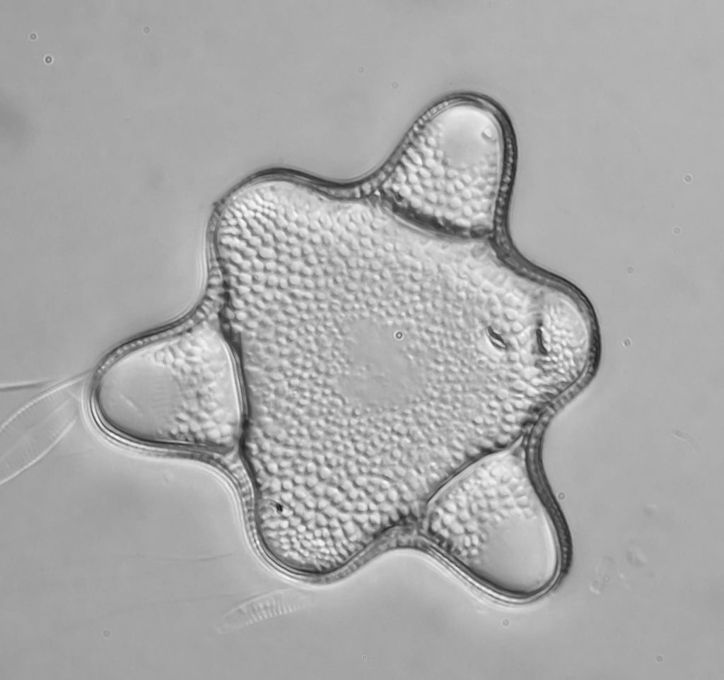
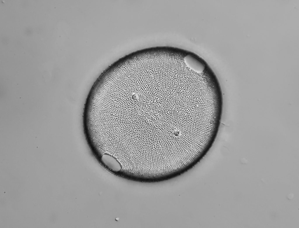
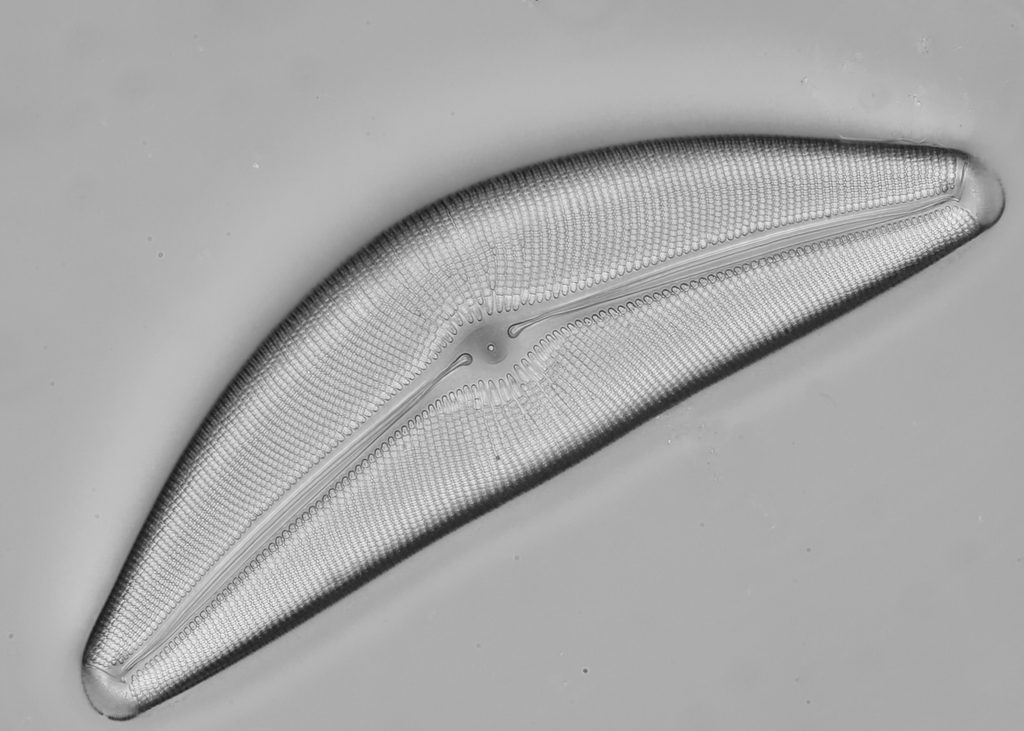
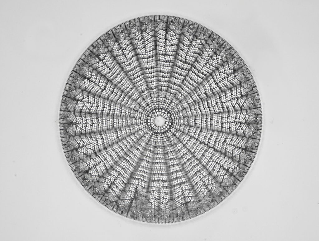
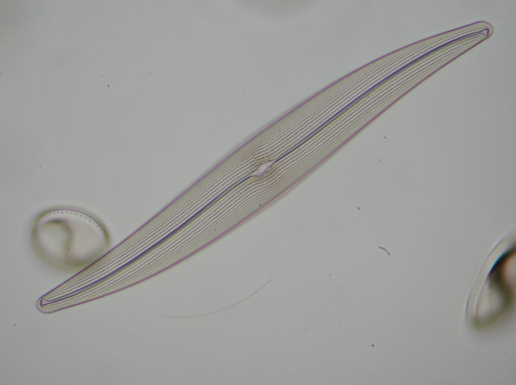
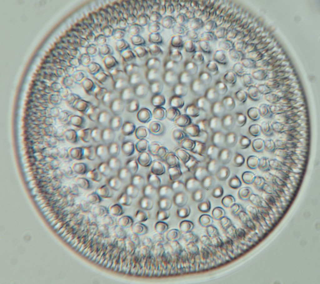
A fossil diatom recovered from diatomite found in what used to be a Japanese lake.
Leave a Reply